
| 015-22 | |
| Regional cerebellar morphometric alterations in early Parkinson’s disease | |
| Bethany Facer | |
| Department of Pharmacology and Therapeutics, Institute of Systems, Molecular and Integrative Biology, University of Liverpool, UK | |
| The Abstract | |
| Abstract Body | Parkinson’s disease (PD) is the second most common neurodegenerative disease primarily characterised by progressive basal ganglia degeneration. Growing evidence suggests areas outside the basal ganglia are also impacted, particularly regions involved in motor functions. We aimed to quantify the volume of individual cerebellar lobules on MRI scans in patients with a new diagnosis of PD to determine cerebellar alterations earlier in the disorder and whether changes relate to principle symptomatic features. PwP had a significant volume increase of the right (t=3.14, pFWE=0.008) and left (t=2.92, pFWE=0.016) cerebellar lobule V, and a strong trend for increased volume of the right VI lobule in patients (t=2.29, pFWE=0.088). There were no significant differences in cerebellar lobule volume between TD and PIGD motor subtypes. |
| Image 1 | 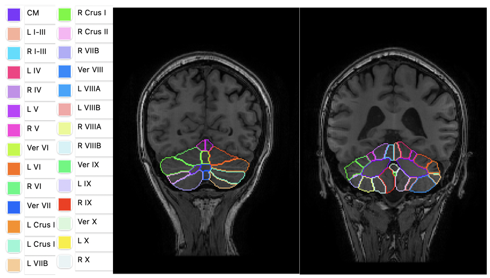 |
| Additional Authors | |
| Christophe de Bezenac | |
| Antonella Macerollo | |
| Thomas Butts | |
| Jibril Farah | |
| Simon Keller | |
| Additional Institutions | |
| The Walton Centre NHS Foundation Trust, Liverpool, UK | |
| School of Medicine, Faculty of Health Sciences and Wellbeing, University of Sunderland, UK |
015-22 – Regional cerebellar morphometric alterations in early Parkinson’s disease
Written by
in